

Researchers in the US have created wearable microscopes to enable unprecedented insight into the signalling patterns that occur within the spinal cords of mice.
This technological advancement, by researchers at the Salk Institute in San Diego, will help researchers better understand the neural basis of sensations and movement in healthy and disease contexts, such as chronic pain, itch, ALS or multiple sclerosis (MS).
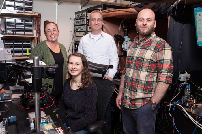
Senior author Axel Nimmerjahn, associate professor and director of the Waitt Advanced Biophotonics Center.
“These new wearable microscopes allow us to see nerve activity related to sensations and movement in regions and at speeds inaccessible by other high-resolution technology.
“Our wearable microscopes fundamentally change what is possible when studying the central nervous system.”
The wearable microscopes are approximately seven- and fourteen- millimetres wide (about the width of the human spinal cord) and offer high-resolution, high-contrast, and multicolour imaging in real-time across previously inaccessible regions of the spinal cord.
This new technology can be combined with a microprism implant, which is a small reflective glass element placed near the tissue regions of interest.
Erin Carey, co-first author of one of the studies and researcher in Nimmerjahn’s lab, said:
“The microprism increases the depth of imaging, so previously unreachable cells can be viewed for the first time.
“It also allows cells at various depths to be imaged simultaneously and with minimal tissue disturbance,” says
Pavel Shekhtmeyster, a former postdoctoral fellow in Nimmerjahn’s lab and co-first author on both studies, added:
“We’ve overcome field-of-view and depth barriers in the context of spinal cord research.
“Our wearable microscopes are light enough to be carried by mice and allow measurements previously thought impossible.”
With the novel microscopes, the research team began applying the technology to gather new information about the central nervous system.
In particular, they wanted to image astrocytes found in the spinal cord because the team’s earlier work suggested the cells’ unexpected involvement in pain processing.
The researchers found that squeezing the tails of mice activated the astrocytes, sending coordinated signals across spinal cord segments.
Before the invention of the new microscopes, it was impossible to know what astrocyte activity looked like—or what any cellular activity looked like across those spinal cord regions of moving animals.
Daniela Duarte, co-first author of one of the studies and researcher in Nimmerjahn’s lab, said:
“Being able to visualise when and where pain signals occur and what cells participate in this process allows us to test and design therapeutic interventions.
“These new microscopes could revolutionise the study of pain.”
The research has already begun investigating how neuronal and non-neuronal activity in the spinal cord is altered in different pain conditions and how various treatments control abnormal cell activity.
Image: Salk Institute